
Figure 2.
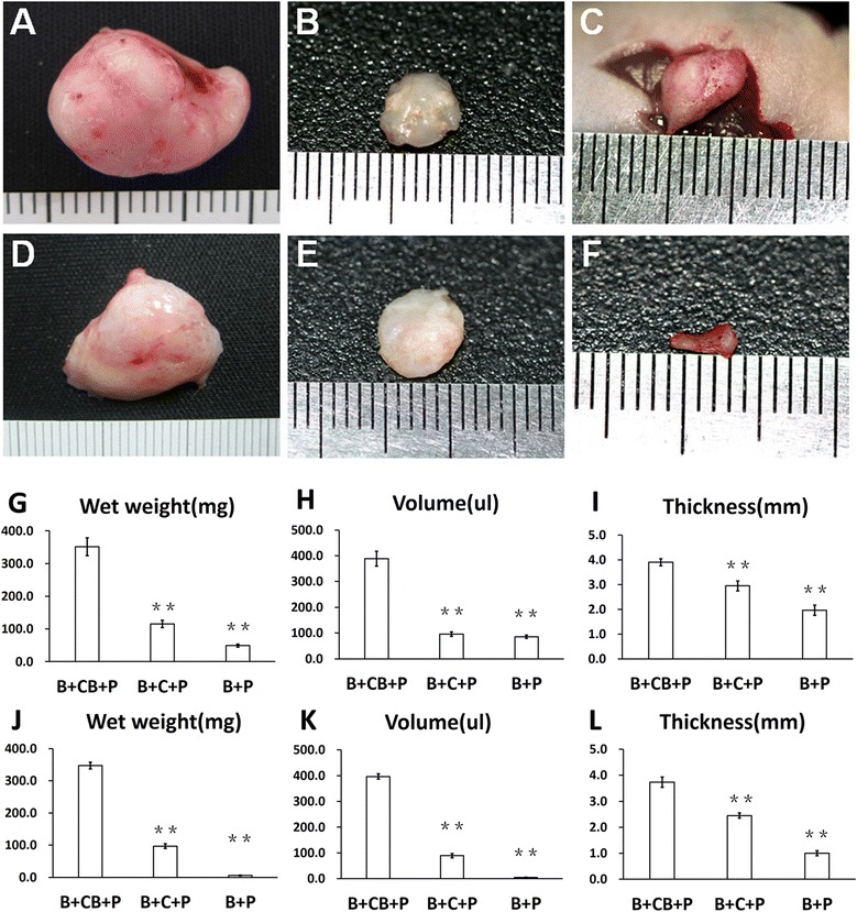
Macroscopic examination of regenerated tissue form grafts. Close macroscopic views of the regenerated cartilage from the B-CB-P (A, D), B-C-P (B, E) and B-P (C, F) groups after 4 and 12 weeks of in vivo incubation. Samples harvested from mice at 4 weeks (G, H, I) and 12 weeks (J, K, L) postoperatively present different volume, weight and thickness. **P <0.01. B-C-P, BMSCs–chondrocytes–PRP; B-CB-P, BMSCs–cell bricks–PRP; B-P, BMSCs–PRP; BMSC, bone marrow-derived mesenchymal stem cell; PRP, platelet-rich plasma.