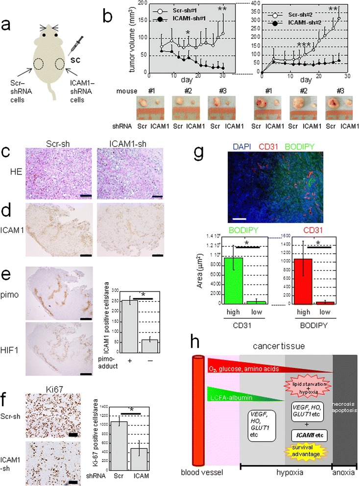
Figure 9.

ICAM1 is inducible in vivo and confers a growth advantage to CCC tumors. a) Sites where NOD-SCID mice were injected with cells expressing Scr-shRNA (#1 or #2) or ICAM1-shRNA (#1 or #2). b) Tumor volumes in mice injected with Scr-shRNA #1 or ICAM1-shRNA #1 (n = 9) or Scr-shRNA #2 or ICAM1 shRNA #2 (n = 10). *P = 0.0006; **P < 0.0001; ***P = 0.002. c) H&E staining (scale bar: 100 μm). Immunohistochemistry of ICAM1 d) Pimo-adduct and HIF1 e). Quantitation of ICAM1-positive cells within pimo (+) or (−) areas (n = 8). *P < 0.0001. Scale bar: 500 μm. f) Immunohistochemistry of Ki67. Scale bar: 100 μm. Quantitation of Ki67 positive cells is shown (n = 8). *P < 0.0001. g) Lipid staining using frozen sections of tumor tissue. Immunofluorescence of CD31 was also performed for vessels. Scale bar: 100 μm. The relationship between neutral lipid staining with BODIPY and CD31 positivity was quantified (n = 10). *P < 0.0001. h) Hypothetical model of lipid starvation and hypoxia associated with induction of genes of different categories in tumor tissues. Diffusion of LCFAs associated with high molecular weight albumin from the blood stream is limited compared to that of smaller molecules (O2, glucose, and amino acids). Unlike (VEGF), (HO) and (GLUT1) genes, Sp1-dependent genes such as ICAM1 may be efficiently up-regulated only under hypoxia with a limited LCFA supply, resulting in a survival advantage of cancer cells to such a severe microenvironment.
