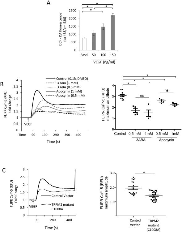
Figure 1. TRPM2 mediates VEGF-induced Ca2+ entry in endothelial cells.
A) DCFH-DA fluorescence measured in confluent human pulmonary artery ECs stimulated with VEGF. VEGF increased the DCF fluorescence in dose- and time-dependent manner. Experiments were independently reproduced three times in quarduplets.
B) Apocynin (Apo) and 3ABA prevent VEGF-induced Ca2+ entry in ECs. ECs were grown to confluence and were pre-treated with apocynin or 3ABA at indicated concentration 2h before loading ECs with FLIPR-5 Ca2+ sensitive dye. VEGF was added at the arrow (60 s). Although, there was a trend for inhibition of Ca2+ influx at 1mM concentrations of apocynin or 3ABA compared to lower doses, the values were not significant (ns). The experiment was repeated twice in quarduplets. Mean of maximum responses is expressed in the scattered dot plot (with SEM).
C) Ca2+ transients measured using FLIPR-5 Ca2+ sensitive dye following VEGF stimulation of ECs transfected with ADPR-insensitive TRPM2 mutant C1008A or empty vector. VEGF in each case was added at the arrow. Ca2+ ionophore ionomycin was added at the end of experiment for calibration. VEGF-mediated Ca2+ influx was suppressed in cells expressing the TRPM2 mutant (Cys1008 substituted to Ala). Representaive tracing is shown. The experiment was repeated twice with each experiment consisting of 5 or more replicates. Mean of maximum responses is expressed in the scattered dot plot (with SEM).

