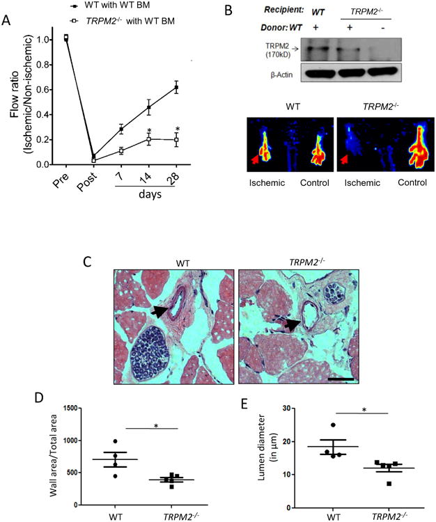
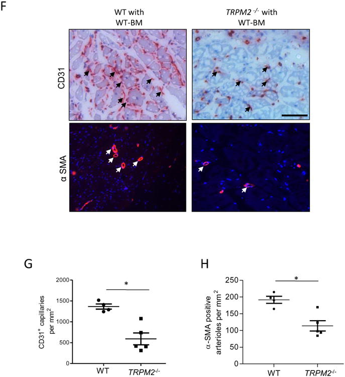
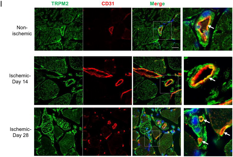
Figure 6. Impaired post-ischemic neovascularization in TRPM2−/− mice.


A) Blood flow recovery after hindlimb ischemia in WT and TRPM2−/− mice shown as ratio of laser Doppler-measured perfusion values (SEM) of ischemic (left) and non-ischemic (right) legs (WT, n = 4; TRPM2−/−, n = 5). Right panel show representative perfusion images at day# 28 measured by laser Doppler.
B) Effects of bone marrow transplantation of WT myeloid cells into WT and TRPM2−/− recipients. Analysis of bone marrow myeloid cells showed effective reconstitution of TRPM2 in TRPM2−/− mice.
C-E) Hematoxylin and eosin (H&E) staining of mouse adductor muscle. Arrowhead shows collateral vessels in the semi-membranosus muscle. The wall area is calculated from measurements of luminal and perivascular tracings. The lumen diameter was calculated for vessels in the range 5-30μm diameter size. Data are shown as scatter plots (with SEM). Bar represents 50 μm.
F-H) Histological analysis of mouse gastrocnemius (GC) muscle showing CD31 and α-SMA immunostaining obtained at day#28 after femoral artery ligation. Staining (red) by both histological and immunofluorescenee show CD31+ capillaries and α-SMA+ arterioles; DAPI stained nuclei are blue. Scale bar represents 50 μm. Scatter plots (with SEM) shows quantification of the number of CD31+ and α-SMA+ structures per mm2 area (*p<0.05).
I) Immunofluorescent staining of GC muscle at days #14 and #28 of post-ischemic injury. Expression of TRPM2 was localized in endothelium of vessels. Ischemia induced the expression of TRPM2 in endothelial cells of newly formed vessels at day#14 and #28 post-induction of ischemic injury. Scale bar represent 20μm.
