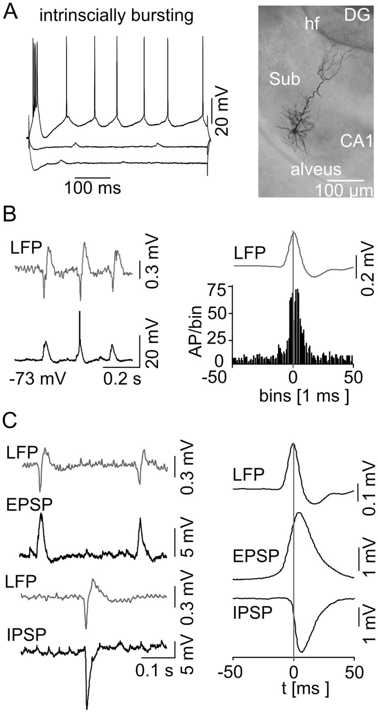
Fig 3. Behavior of subicular IB cells during spontaneous subicular SPWs.

(A) Current-voltage relationship of a IB cell (left) with current injection steps of −300 pA, −100 pA, and +140 pA, respectively, displayed with the attached microphotograph of a biocytin-stained IB cell (right). These cells exhibit the typical pyramidal shaped cell body, prominent apical dendrites that travel through the molecular layer reaching the hippocampal fissure (hf), and basal dendrites that spread within the pyramidal cell layer. The axon leaves the subiculum (Sub) via the alveus. IB cells respond to a hyperpolarizing current injection with a sag in membrane potential whereas a positive current pulse leads to burst firing. (B) Example of simultaneous extracellular LFP (top trace) and intracellular (bottom trace) recordings at RMP is shown on the left. The intracellular recording reveals phase-locked synaptic responses as well as a full-blown AP (truncated for clarity) with respect to the LFP SPWs. The spike time histogram (n = 16 IB cells) on the right illustrates a clear peak of AP generation in close vicinity to the SPW peak. The vertical line marks the maximum mean SPW deflection as time point 0. (C) EPSPs and IPSPs are displayed in correlation to the LFP (left). The EPSPs and IPSPs were recorded at −80 mV and at 0 mV, respectively. (right) Accumulated mean EPSP/IPSP with respect to the maximum SPW peak deflection.
