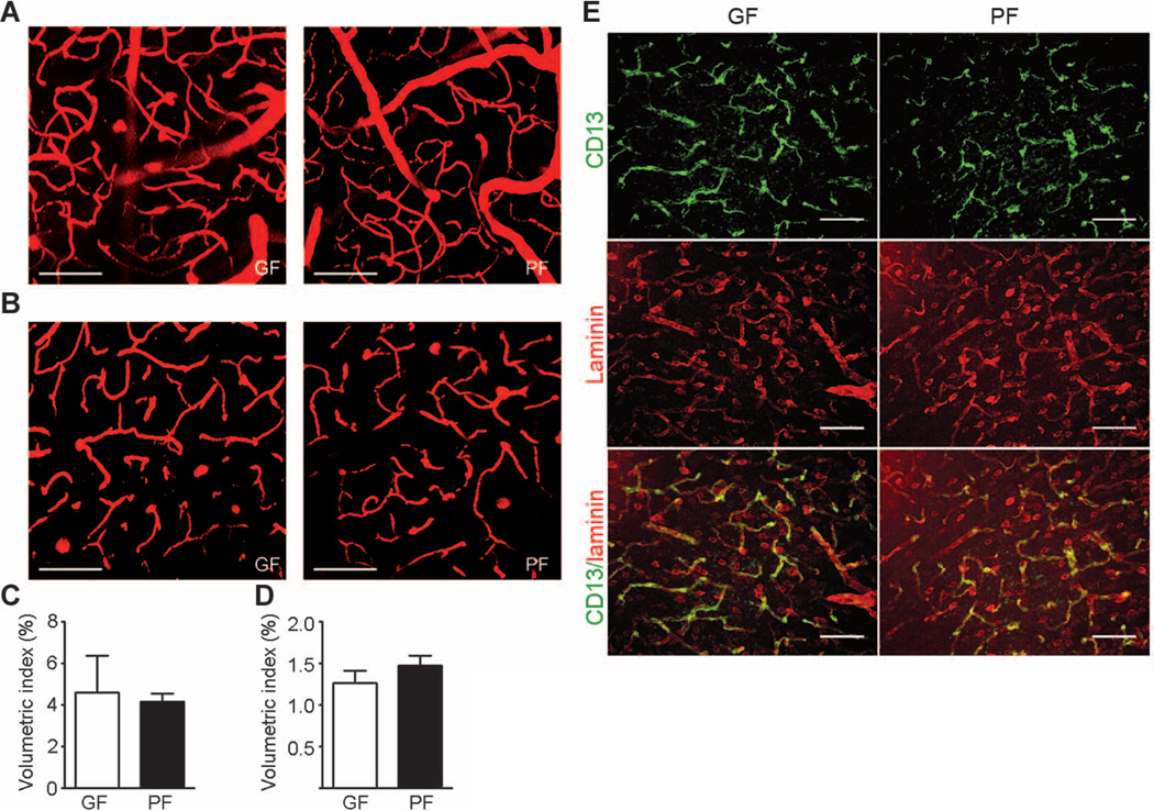
Fig. 3. Brain blood vessel and pericyte coverage in germ- and pathogen-free adult mice.
(A to D) Two-photon imaging of brain blood vessels in germ-free (GF) and pathogen-free (PF) adult mice. Tetramethyl rhodamine isothiocyanate (TRITC)–dextran was applied retro-orbitally to highlight the brain blood vessels. (A) Representative images of brain vasculature 20 to 80 µm below the dura mater in germfree (left panel) and pathogen-free (right panel) mice reveal mainly large vessels (average diameter, ~40 µm). (B) Representative images of the brain vasculature 120 to 180 µm below the dura mater in germ-free (left panel) and pathogen-free (right panel) mice showing mainly capillaries. (C) Quantitative analysis of blood vessel density 20 to 80 µm below the dura mater. (D) Quantitative analysis of blood vessel density 120 to 180 µm below the dura mater. Scale bars, 100 µm. Data are representative of n = 3 independent experiments. (E) Representative images of pericyte coverage (CD13, green) in the cerebral cortex of pathogen- and germ-free mice (n = 4 mice per group). Laminin (red) was used as an endothelial cell marker. Scale bars, 50 µm.

