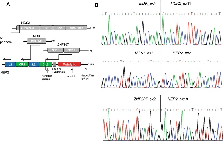
Figure 1.

Identification of HER2 fusions in gastric cancer. A. Schematic illustration of the wild-type HER2 protein and the three fusions identified in this study. The breakpoints for each fusion are indicated by arrows. B. Sanger sequencing of the fusion junctions.
