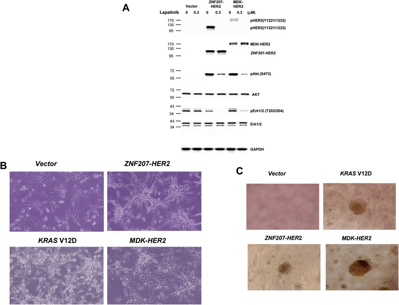
Figure 5.

Transformation of NIH/3T3 cells by ZNF207-HER2 and MDK-HER2 fusions. A. Modulation of HER2 signaling in NIH/3 T3 cells expressing HER2 fusions. Cell lysates collected from cells stably expressing HER2 and HER2 fusions were subjected to western blotting analysis with antibodies against phospho and total HER2, Akt and Erk. B. Exogenous expression of ZNF207-HER2 and MDK-HER2 led to transformational morphological changes. Cells were photographed under a phase-contrast light microscope (×150) under identical conditions. C. Anchorage-independent growth of NIH/3 T3 cells expressing HER2 fusions. Cells were seeded in soft agar culture in 96-well plates for 14 days and colony formation was photographed under a phase-contrast light microscope (×150). The results are representative from three independent experiments.
