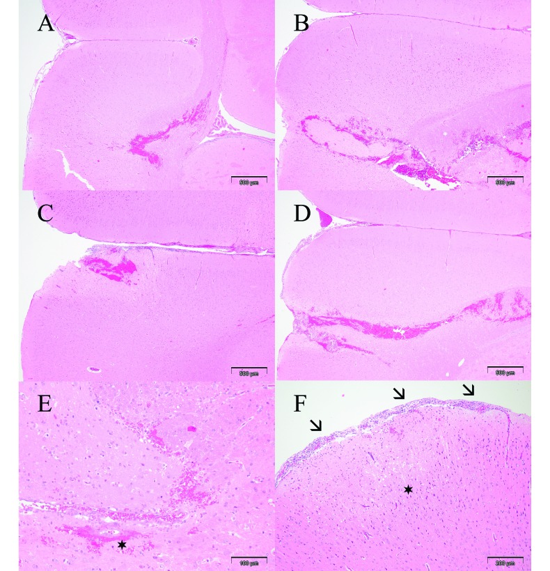
Figure 2.
Coronal brain sections (magnification, 4×) showing intracerebral injection channel in rats treated (A) with vehicle only or (B) 7.96 × 10⁵ pfu, (C) 7.96 × 10⁶ pfu, or (D) 7.96 × 10⁷ pfu H1PV. (E) Parenchymal hemorrhages and focal minimal vasculitis (asterisk) in a rat treated with vehicle only (magnification, 20×). (F) Mild meningeal inflammatory cell infiltrates (arrows) and a small area of encephalomalacia (asterisk) near the injection channel in a rat treated with 7.96 × 10⁶ pfu H1PV (magnification, 10×).

