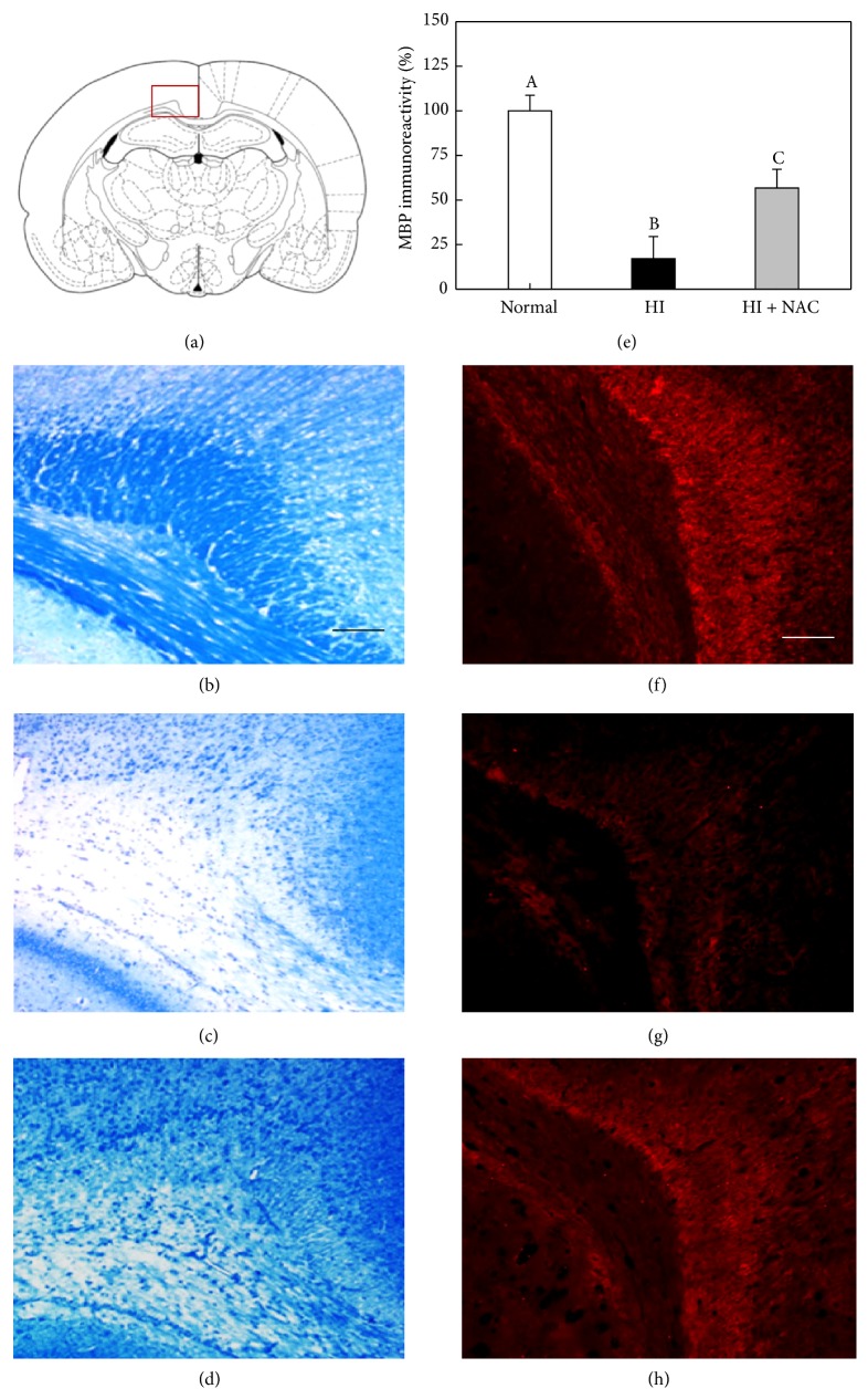
Figure 5.
Recovery of LFB-stained myelins and MBP immunoreactivity in rat corpus callosum by NAC. Integrity of myelins was analyzed with LFB staining and MBP immunoreactivity at postnatal day 45. The square box in (a) represents an examined area of corpus callosum. The brain tissues were stained with LFB (b–d) or MBP antibody (f–h). (b, f) Normal animals, (c, g) HI alone, and (d, h) HI + NAC. Scale bars = 500 μm. Values with different superscript letters in (e) represent a significant difference in MBP immunoreactivity from (f–h), P < 0.05.

