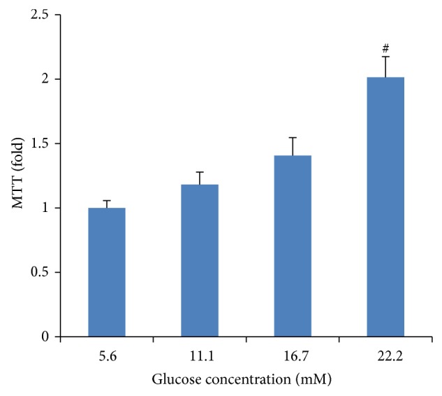
Figure 3.

High-glucose-induced proliferation of vascular smooth muscle cells (VSMCs). VSMCs were incubated in various concentrations of glucose (5.6, 11.1, 16.7, and 22.2 mM) for 48 h. Cell proliferation was measured by an MTT assay. Mannitol was used as an osmotic control. Data expressed as mean ± SEM. ∗ P < 0.05 versus normal glucose (5.6 mM) group; # P < 0.01 versus normal glucose (5.6 mM) group.
