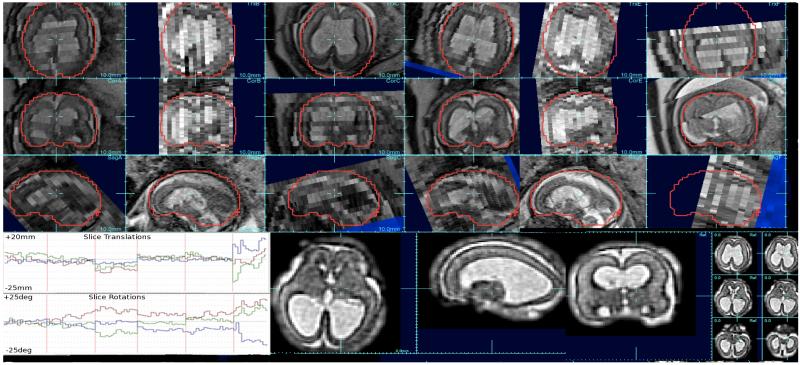
Figure 1.
Example clinically acquired multi-slice study (upper 3 rows) where 6 stacks (A-F) of 1×1mm slices with thicknesses varying between 3mm and 4mm were acquired of a fetus with enlarged ventricles: note the motion between odd and even slices (data acquired with an interleave of 2), differences in slice intensity due to through plane motion and intensity bias, and low through-plane resolution. Between slice motion was estimated using SIMC (Kim et al., 2010) (see bottom left traces of rotation and translation corrections of each slice pose) and signal differences were corrected using SIBC ((Kim et al., 2011). A 3D image reconstruction was created using a robust iterative scheme with iterative slice profile deconvolution (Fogtmann et al., 2012) to create a 1×1×1mm resolution voxel size (bottom right).

