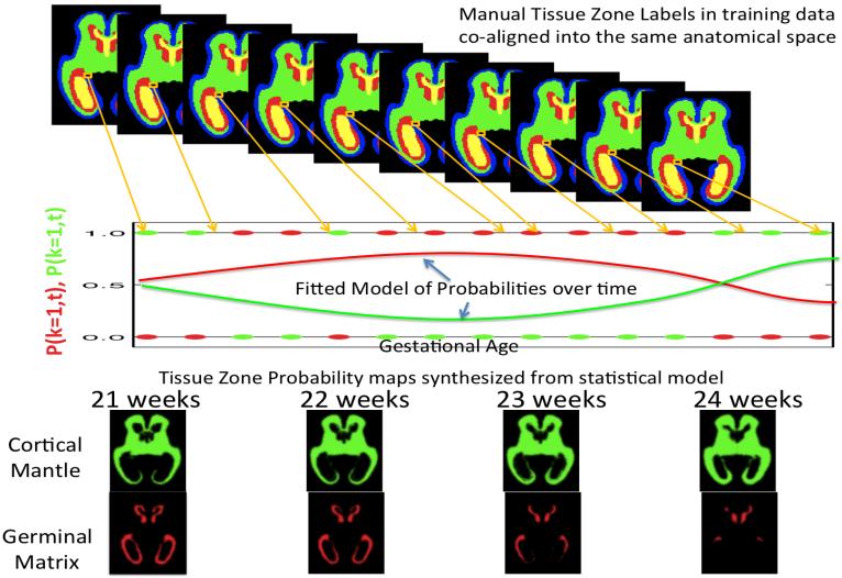
Figure 2.
Using Manual Tracing data to learn the probability of developing tissues zones visible in MRI scans of fetuses of different ages: For each voxel, each manually marked dataset mapped to a common fetal coordinate system contributes a binary decision at a given age (red and green dots on graph). A constrained mathematical model is fitted to these binary observations to form a continuous estimate of the probability of observing a tissue class (red and green curves) at any given age at that point in the anatomy. The formation of such a complete computational model that also captures age related changes in fetal brain shape and appearance (contrast) in MRI allows the formation of age specific templates and tissue priors for any given gestational age (bottom row). These age specific templates can be used as a starting estimate for automated techniques that label tissues in new MRI scans (Habas et al., 2010a) by adapting the average model to the new scan of an individual.

