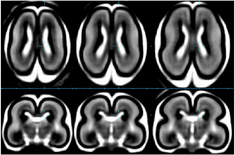
Figure 4.
Average brain tissue contrast and shape captured at 20GW (left), 21GW (middle) and 22GW (right) in a T2W MRI growth model (Habas et al., 2010a) derived from in-utero data of 40 healthy subjects. Outer surface consists of dark cortical plate below which a bright region of subplate is visible. Around the ventricles is the dark region of germinal matrix.

