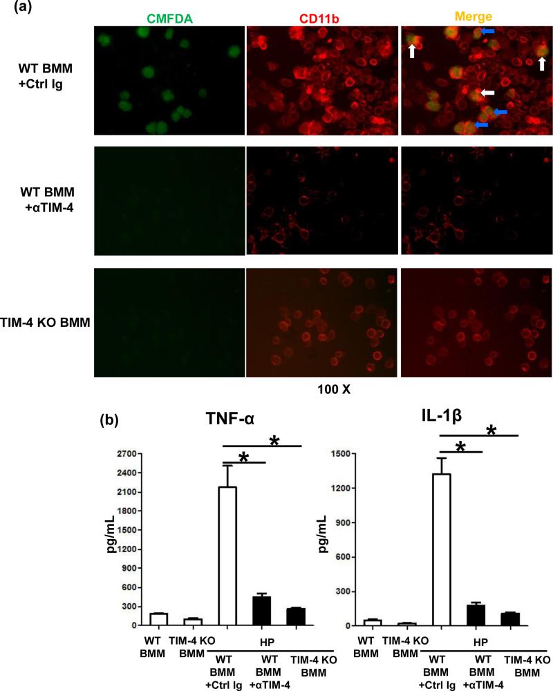
Figure 7.
TIM-4 signaling in macrophage phagocytosis/activation in vitro. (a) CMFDA-stained H2O2-necrotic hepatocytes (green) co-cultured with CD11b+ BMMs (red) from WT + control Ig vs. TIM-4 mAb treatment; or from TIM-4 KO donor mice. “White” and “blue” arrows represent captured and engulfed hepatic debris by BMMs, respectively. (b) ELISA-assisted TNF-α and IL-1β detection in co-culture supernatants (*p<0.001). Representative of n=3/group.

