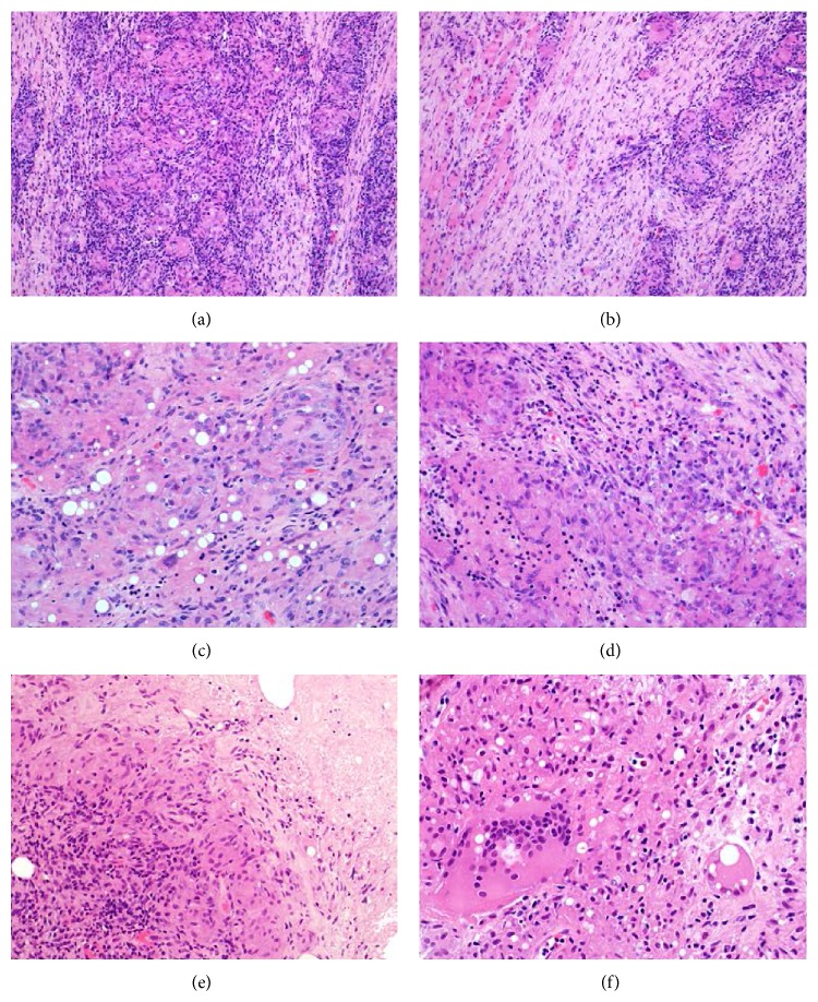
Figure 3.
(a) These are relatively ill-defined granulomas comprising sheets of epithelioid histiocytes and giant cells and are surrounded by prominent mixed inflammation rich in small lymphocytes. (b) This example occurred deeply within the biceps muscle. There is atrophy of skeletal muscle fibers, with surrounding fibrosis and chronic inflammation. (c) In areas, there are rounded clear intracytoplasmic vacuoles within the histiocytic population. This is thought to represent foreign body reaction to polylactic acid. Luteinising hormone-releasing hormone (LHRH) has been shown to have lipolytic activity in vitro, and the degenerate lipid granules may induce foreign body granulomas. (d) The chronic inflammatory infiltrate comprises largely histiocytes and small lymphocytes, but there are smaller numbers of plasma cells and eosinophils. (e) This example shows areas of necrosis. (f) The giant cells can contain twenty or thirty nuclei. Engulfment of vacuoles corresponding to degenerate elements of leuprorelin acetate is apparent.

