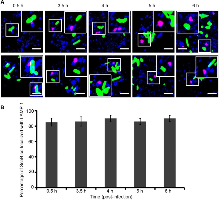
Fig 8. As SseB moves away from the Salmonella surface, it is associated with the vacuolar membrane.
(A) RAW264.7 macrophages were infected with WT Salmonella harboring pFPV25.1 for the constitutive expression of GFP (green). Cells were fixed, permeabilized, and immunostained for mouse monoclonal anti-LAMP-1 (blue) and rabbit polyclonal anti-SseB (red) followed by Alexa568- or Alexa647-labeled secondary antibodies, respectively, for various times as indicated. The merged image indicates that once SseB was detached from the bacterial cell surface, it was co-localized with the host endosomal membrane (i.e., LAMP-1–positive membranes). Images were obtained using the Nikon A1R confocal microscope. Localization resulted in magenta staining of Salmonella SseB as analyzed by Image J software. Scale bar = 3μm. (B) Results of three independent experiments in which macrophages were infected with WT GFP-expressing Salmonella and immunostained with LAMP-1 and SseB at 3.5 h, 4 h, 5 h, and 6 h post-infection. The values represent the mean ± SEM.

