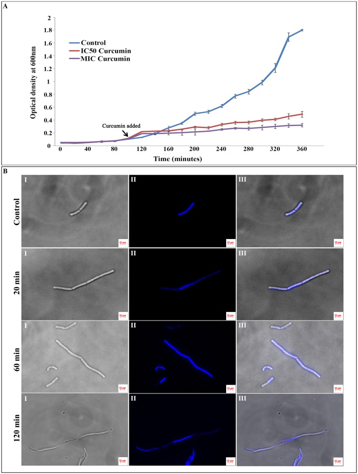
Fig 1. Effect of curcumin treatment on the B. subtilis AH75 growth and cell morphology.
(A) B. subtilis AH75 strain was grown in LB media having spectinomycin antibiotic (100 μg/mL) till the OD600 reached to 0.1. Then the cultures were treated with DMSO (control), 20 μM (IC50 concentration) and 100 μM (MIC concentration) curcumin. Growth curve was plotted by measuring the OD600 for all the samples at every 20 min interval till 360 min (mid-exponential phase). The three time points of curcumin treatment (20, 60 and 120 min) used in proteomic analysis are indicated by arrows. IC50 concentration was used for subsequent proteomic analysis. (B) B. subtilis AH75 strain was grown in the presence of 20 μM (IC50 concentration) curcumin and the samples was collected after 20, 60 and 120 min of the drug treatment. Cultures treated with only DMSO was used as control. The nuclear materials were stained using 1 μg/μL DAPI for 20 min at room temperature in dark for all the samples. The fluorescence microscopic images were captured with both DAPI and DIC filters. The control B. subtilis cells showed normal cell length with one or two nucleoids per cell whereas after 20, 60 and 120 min of incubation with 20 μM (IC50 concentration) curcumin, most of the cells turned into filamentous structure with multiple nucleoids. I- DIC image, II- DAPI image and III- overlay image.

