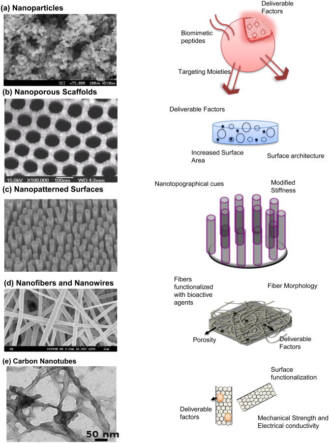
Figure 1. Nanomaterials in tissue engineering.
(a) (Left) SEM of ZnO nanoparticles. Reprinted with permission from Ref. 44 Copyright 2011 Springer (Archives of Toxicology); (Right) Schematic of a functional nanoparticle, which provides a reliable conduit for delivering drugs, growth factors, cytokines and other factors. These can be functionalized with biomimetic peptides, targeting moieties and functional groups for degradation in response to appropriate physiological stimuli. (b) (Left) SEM image of nanoporous alumina. Reprinted with permission from Ref. 69 Copyright 2007 Elsevier Ltd. (Biomaterials); (Right) Schematic of a nanoporous scaffold. These scaffolds make for a three dimensional construct with increased surface area, surface nanoarchitecture and an ability for controlled delivery of factors. (c) (Left) SEM image of metallic glass nanopillars. Reprinted with permission from Ref. 39 Copyright 2014 ACS Nano ; (Right) Shematic of nanopatterned Surfaces, which present nanotopographical and biomechanical cues to engineer cellular response to tissue engineering scaffolds. (d) (Left) SEM image of PLGA/Collagen nanofibers. Reprinted with permission from Ref. 90 Copyright 2010 Elsevier Ltd. (Journal of Membrane Science); (Right) Schematic of a nanofibrous scaffold. Nanofibers and nanowires contribute to creation of biomimetic scaffolds that recreate the native extracellular matrix structure. These can also be functionalized/loaded with drugs and other factors for controlled localized delivery. (e) (Left) TEM image of a gelatin-methacrylate coated nanotube. Reprinted from Ref. 24 Copyright 2013 ACS Nano.; (Right) Schematic of carbon nanotubes. These are used to reinforce mechanical strength and promote electrical conductivity in polymers and hydrogels used in tissue engineering applications. They can also serve as excellent drug delivery vehicles.

