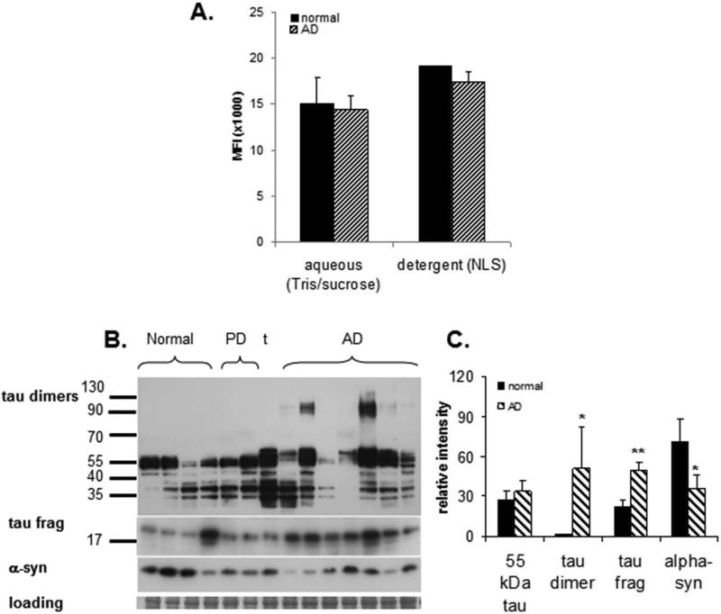
Fig. 2. Biochemical analysis of tau in synaptosome-enriched fractions (SEF, P-2, crude synaptosomes).
(A) Distribution of tau in sequential buffer and detergent (1% N-laurylsarcosyl) extracts measure by bead-based immunoassay: MFI=mean fluorescent intensity (B) Western blot analysis of tau peptides (HT7 antibody) in a series of aged normal control (N = 4) and AD samples (N = 7). Controls include 2 Parkinson's disease cases and 1 tauopathy case (t); the right PD case (lane 6) had a 2-year history of dementia; case information is presented in Table 2 (C) Quantification of Western blot in B; *p<0.05, ** p<0.01.

