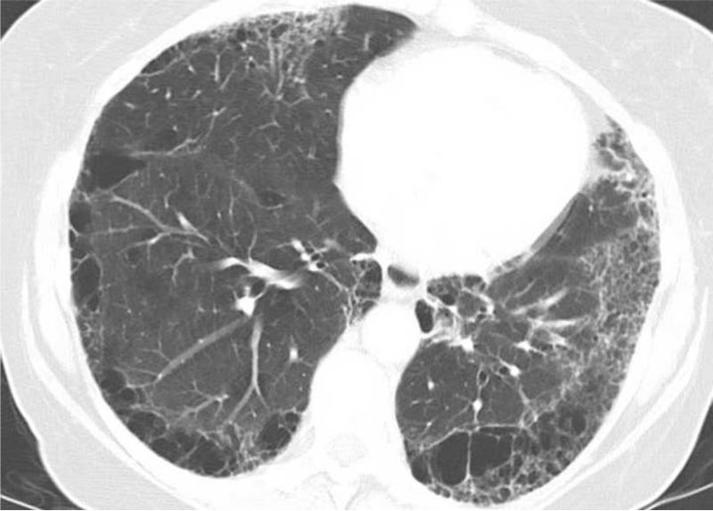
Fig. 5.
62-year-old, female with stage IVB NSCLC with 27 pack year history of smoking.
Baseline chest CT at diagnosis demonstrated bilateral fibrosis in multiple lobes with honeycombing and traction bronchiectasis in subpleural distribution (a, b), which was scored as 3 (ILA). The mass in the left lower lobe represents the primary tumor (a). Also note underlying emphysema. The patient was subsequently treated with systemic therapy using carboplatin and pemetrexed. The patient experienced progressive disease and significant increase of dyspnea over the course of 6 months, with oxygen saturation of 82% on room air, requiring 3 liters of oxygen at rest. The overall survival since the diagnosis of NSCLC was 6.5 months.

