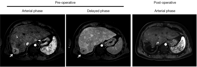
Figure 1.

Dynamic contrast MRI. Pre-operative scans show a hypervascular mass in segment 7 on arterial phase, followed by delayed washout (arrows). A small hypervascular lesion without definitive washout in the caudate lobe can also be visualized (arrowhead). Corresponding post-operative arterial phase MRI shows surgical absence of lesions in segments 7 and 1.
