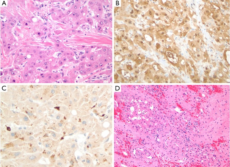
Figure 3.
Fibrolamellar HCC. Histologic findings highlight (A) neoplastic cells arranged in cords and trabecular, intermixed with unpaired arteries and thick fibrous strands (H&E, 100×); (B) arginase-1 and (C) CD68 expression based on immunohistochemical staining of neoplastic cells (100×); and (D) surrounding liver tissue showing sinusoidal dilatation, hemorrhage, hepatocytic dropout and fibrosis in the centri-zonal region (100×). HCC, hepatocellular carcinoma.

