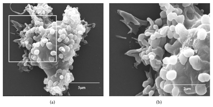
Figure 3.

Scanning electron microscopy of anti-Gal coated α-gal nanoparticles binding to macrophages via Fc/FcγR interaction. α-gal nanoparticles coated with anti-Gal Ab were coincubated with adherent GT-KO pig macrophages for 2 hours at room temperature, washed to remove nonadherent nanoparticles, and processed for electron microscopy analysis. Many α-gal nanoparticles adhere to the surface of a representative macrophage. The inset in (a) is enlarged in (b). Modified from [23].
