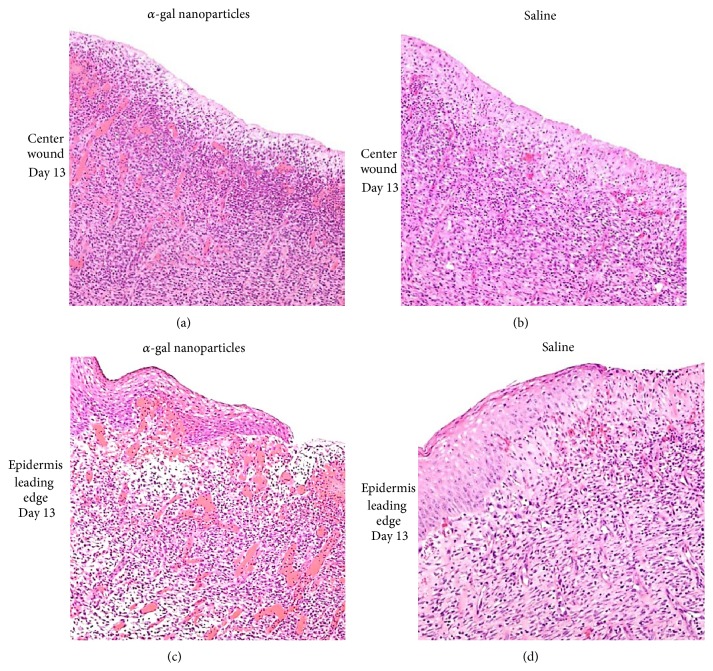
Figure 4.
Vascularization of GT-KO pig wounds treated with α-gal nanoparticles (100 mg) or with saline and studied on day 13. The presented histology is of the centers of wound (not covered by regenerating epidermis), or wound areas under the leading edge of regenerating epidermis treated with α-gal nanoparticles (a and c, resp.) or with saline (b and d, resp.). There are many more macrophages and blood vessels (filled with red stained RBC) in wound treated with α-gal nanoparticles than in those treated with saline. Representative wounds from 6 GT-KO pigs (H&E ×200). Modified from [21].

