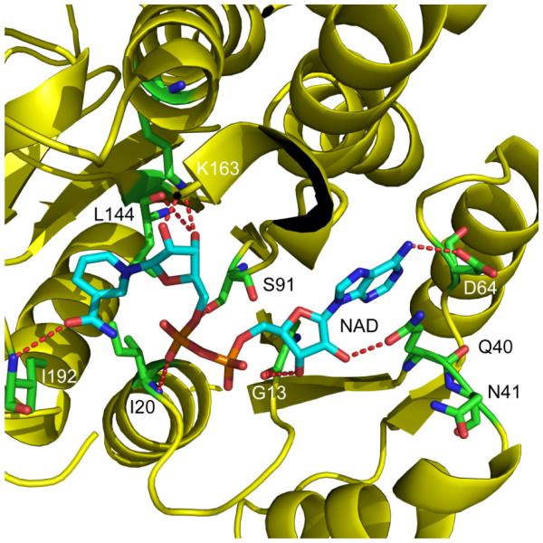
Figure 1. Structure of the ecFabI:NAD+ Complex.
Structure of ecFabI complexed with NAD+ (pdb code 1DFI) showing interactions between the protein and the NAD+ ribose. ecFabI is colored yellow and polar interactions between the protein residues (green) and NAD+ (cyan) are indicated with red dashed lines. Q40 interacts with the 2’-hydroxyl group in the adenosine moity of NAD+. The Figure was made using pymol (44).

