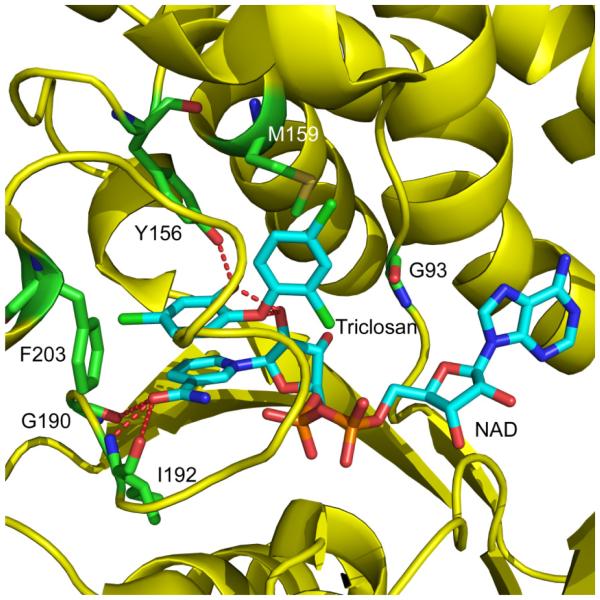
Figure 3. Structure of ecFabI Complexed with NAD+ and Triclosan.
Structure of triclosan bound to ecFabI (pdb code 1D8A) showing the proximity of residues G93, I192 and F203 to the inhibitor binding site. The corresponding residues in saFabI were found to be mutated in the diphenyl ether resistant S. aureus strains. ecFabI is colored yellow, while the polar interactions between the residues (green) in ecFabI and NAD/triclosan (cyan), are indicated with red dashed lines. The Figure was made using pymol (44).

