
Figure 4.
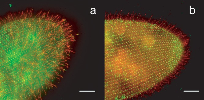
(a) Wild type Paramecium cell with the FLAG-epitope-tagged small-conductance calcium ion–activated potassium ion channel SK1a. The FLAG-tagged channel is visualized with an anti-FLAG primary and a red fluorescent secondary antibody. The green fluorescence of the cell body and spots in the cilia are from an antibody against the folate chemoreceptor and secondary antibody. Note the red cilia, which indicate that the SK1a channel localizes primarily to the cilia. (b) Wild type Paramecium cell with the PKD2 channel FLAG-epitope tagged. The red fluorescence indicates that this channel is present in cilia and also on the general cell surface. The green punctate fluorescence on the cell body is from the primary and secondary antibodies used to identify centrin, a protein known to be present in basal bodies at the base of the cilia. The scale bar indicates 10 micrometers. Source: Reprinted with permission from Valentine and colleagues (2012).