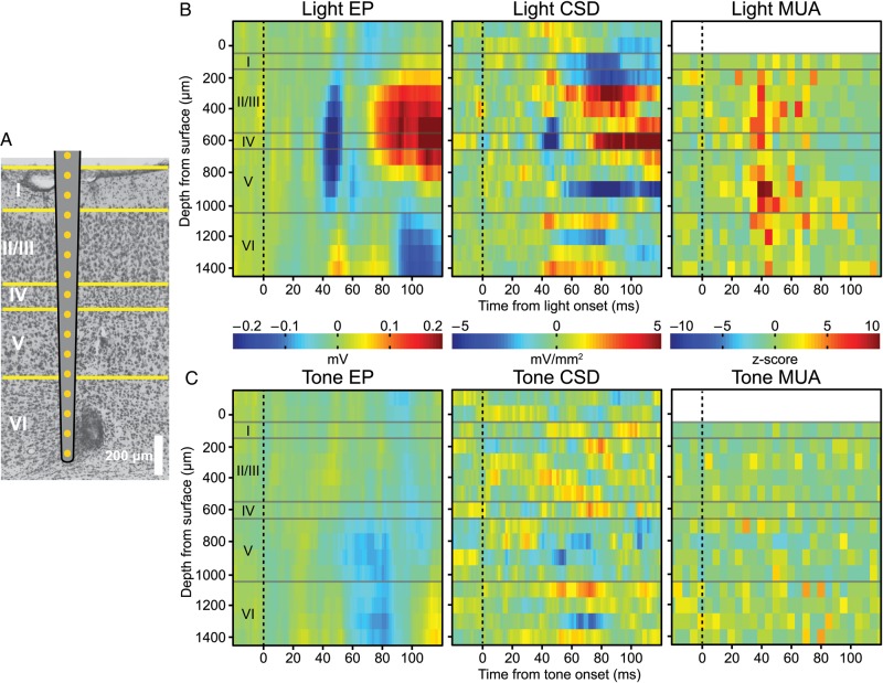
Figure 4.
Tone-evoked EPs in V1 were strongest in infragranular layers. (A) Laminar silicon electrode arrays that encompassed all cortical layers were implanted in V1. Histological validation of electrode placements were performed on Nissl stained sections. A marking lesion was placed on the second to last electrode. (B) Presenting the light produced an EP centered on Layer 4 with an ∼45 ms latency. CSD revealed that the generator of the EP was localized to Layer 4, while the distribution of MUA spanned all layers except Layer 1, which is cell sparse. (C) Tones produced an EP limited to infragranular layers, with current sinks localized to infragranular layers. No substantial changes in MUA occurred to the tone in any of the layers.

