
Figure 1.
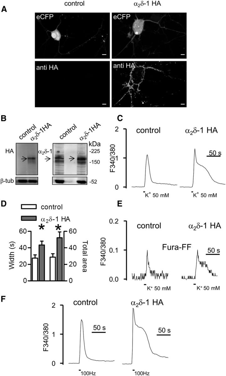
α2δ-1 HA overexpression modulates Ca2+ response duration in DRG neuron cultures. A, 3D projection from z-stacks of confocal images of a DRG neuron transfected with α2δ-1 HA and eCFP (right) or eCFP and nonfunctional Kir2.1-AAA cDNAs (left). Exogenous α2δ-1 HA proteins were detected at the cell surface by live labeling (n = 6 experiments). No staining for the HA epitope was observed in neurons expressing eCFP only. Scale bars, 10 μm. B, Western blotting of neuronal lysates of eCFP or α2δ-1 HA transfected DRGs. Lysates were loaded in duplicate and the membrane was probed for HA (left) or α2δ-1 subunit expression. The neuronal content was quantified by β tubulin III (β-tub) staining (n = 8 DRG cultures, 4 gels). C, Fura-2 imaging of high K+-evoked Ca2+ transients performed in control (left; representative of n = 19) and α2δ-1 HA overexpressing neurons (right; representative of n = 24). Traces are shown after baseline subtraction. D, Control, open bars; α2δ-1 HA, gray bars. The width and total area of transients were significantly higher in α2δ-1 HA overexpressing neurons with respect to control DRGs (*p = 0.01, t test). E, Representative Fura-FF transients imaged in control (n = 11) and α2δ-1 HA neurons (n = 7). F, Examples of Ca2+ transients induced by field stimulation (100 Hz, 10 s) of control (left) and α2δ-1 HA DRGs (right).