Figure 1.
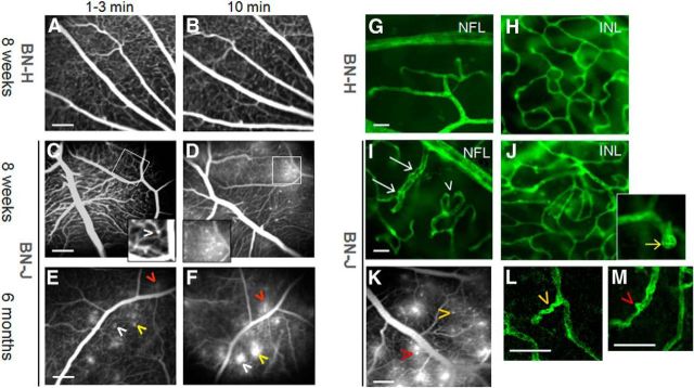
Vascular abnormalities in BN-J rats. Shown is in vivo fluorescein angiography of retinal vessels of BN-H and BN-J rats (A–F). Normal retinal vessels of BN-H rat at early (1–3 min, A) and late phase (10 min, B) of the angiographic sequence. Eight-week-old BN-J rat exhibits subtle capillary dilation hardly detected in the early phase (1–3 min) of angiography (C, arrowhead in the inset) that becomes more visible with leakage at later time point of 10 min (D and inset). At 6 months of age, similar but leakier capillary ectasia are observed (E, F, arrowheads, the same color indicates the same spot). Hyperfluorent leaking dots are observed at 1–3 min and their size increases at 10 min. Scale bar, 200 μm. Confocal imaging of lectin-stained retinal vessels on flat-mounted retinas from BN-H and BN-J rats is shown (G–J, L, M). Normal retinal vascular network (green) at the nerve fiber layer (NFL) and in the deep plexus at the INL from BN-H rat (G, H). In the BN-J rat retina, irregular vascular diameter (white arrows) and increased tortuosity (arrowhead) are observed at the NFL level (I). In the INL, disorganized capillary plexus is observed (J), together with multiple capillary telangiectasia (inset, yellow arrow). Images of a lectin-labeled flat-mounted retina of BN-J rat are linked to their corresponding angiographic pattern (K). Higher magnifications of the lectin-labeled vessels show that leaky telangiectasia (K) correspond to capillary tortuousness (L) and focal capillary ectasia (M). Arrowheads of the same color indicate the same spot. Scale bars: G–J, 20 μm; K, 200 μm; L, M, 50 μm.

