Figure 2.
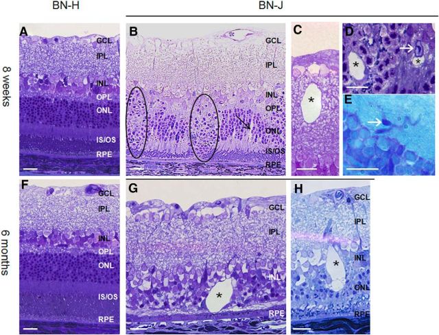
Retinal morphology of BN-H and BN-J rats at 8 weeks and 6 months. Compared with the normally developed retina of BN-H rat at 8 weeks (A), the retina of the BN-J rat shows focal disorganization of the outer retinal layers (B, dark circles), where segments are not formed and nuclei of photoreceptors dive toward the retinal pigment epithelium (RPE). In areas where segments are present, swollen RMG cells can be observed (B, black arrow). Cysts (asterisks) can be found in both the inner (C) and the outer (D) retina. Telangiectasia are also identified on histological sections (D, E, white arrow). At 6 months, the BN-H rat retina is unchanged (F), whereas the retina of the BN-J rat shows variable degrees of degeneration. Photoreceptors have totally disappeared in some areas (G) and cysts are more abundant with irregular shapes (G, H, asterisks). GCL, Ganglion cell layer; IPL, inner plexiform layer; OPL, outer plexiform layer; IS/OS; inner and outer segments of photoreceptors. Scale bar, 20 μm.

