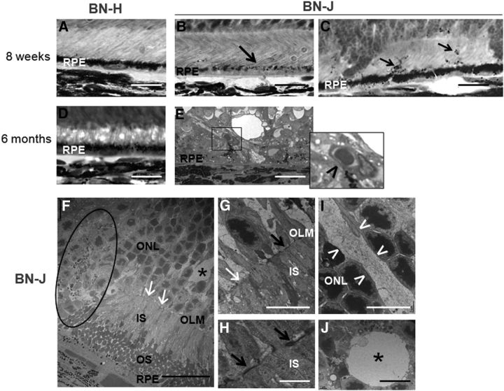
Figure 3.
Outer retinal alterations in BN-J rats. A–E, Histological sections of the outer retina. F–J, TEM images. Contrasting with the heavy pigments located in the apical side of retinal pigment epithelium (RPE) in BN-H retina (A), melanosomes are poorly formed in BN-J rat at 8 weeks, even in areas where the segments have formed (B, black arrow) and pigments migrate in the photoreceptor segment layer (C, black arrows). At 6 months, the outer retina of BN-H rat does not change (D), whereas abnormal vessels are observed between RPE cells and the degenerated retina of BN-J rat (E, inset and black arrowhead), potentially corresponding to neovascularization. TEM analysis allows detection of more subtle changes in BN-J retina such as focal decrease in junction structures (F, G, white arrows) at the outer limiting membrane (OLM), alternating with normal OLM structures (G, H, black arrows). Abrupt disorganization of retinal layers is observed (F, dark circle). Swollen RMG cells (I, in between the white arrowheads) are identified in the ONL and cysts (F, J, asterisks) are surrounded by a membrane-like structure, suggesting intracellular swollen. IS, Inner segments of photoreceptors; OS, outer segments of photoreceptors. Scale bars: A–E, 20 μm; F, 25 μm; G, I, J, 10 μm; H, 2 μm.

