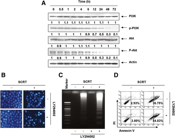
Figure 7.

SCRT triggers apoptosis through an inactivation of PI3K/Akt signaling in A549 cells. (A) The cells were treated with SCRT (1.5 mg/ml) for the indicated times. Equal amounts of cell lysate were resolved by SDS-polyacrylamide gels, transferred to nitrocellulose membranes, and probed with the anti-p-PI3K, anti-p-Akt, anti-PI3K and anti-Akt antibodies. The proteins were visualized using an ECL detection system. Actin was used as an internal control. The relative ratios of expression in the results of the Western blotting were presented at the bottom of each of the results as relative values of the actin expression. (B-D) The cells were pretreated with PI3K inhibitor (LY294002, 50 μM) for 1 h and then treated with SCRT (1.5 mg/ml) for 72 h. (B) After staining with DAPI solution, the nuclei were observed under a fluorescent microscope (original magnification 400x). (C) The genomic DNA from cells was extracted, separated by agarose gel electrophoresis, and visualized under UV light after staining with EtBr. (D) The percentages of apoptotic cells (annexin V+ cells) were analyzed using flow cytometric analysis. The data is the mean of the two different experiments.
