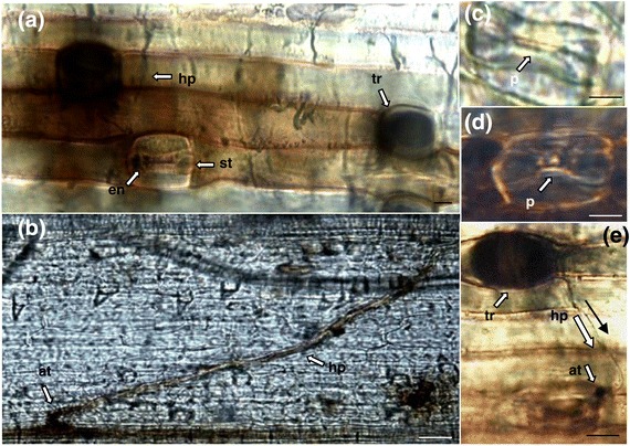
Figure 3.

Light microscopy analysis of leaves of the B. distachyon ecotype Bd21-1, cleared and stained with Trypan Blue and DAB. (a) Fungal hyphae visible on leaf surface (hp), with H2O2 released from cells surrounding a stomata (st), Trypan blue staining is localised in the pore of the stomata (en, st) indicating the presence of fungal tissue. The bases of trichomes (tr) also stain heavily for necrotic tissue. (b) Hyphal attachment (at) at the junction between adjacent vascular tissues showing H2O2 production within the hyphae (hp) and at the site of attachment. Scale bars (a and b) = 0.5 mm (c) Uninfected stomata with clear pore (p) and no evidence of fungal staining. (d) Stomata with clear pore (p) and has not stained for fungal tissue, however H2O2 is present around the circumference of the stomata. (e) Fungal hyphae (hp) originating from the base of a trichome (tr), attaching to the periphery of the stomata (at). Scale bars (c-e) = 0.1 mm
