
Figure 1.
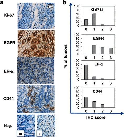
Immunohistochemical expression of Ki-67, EGFR, ER-α and CD44 in canine mammary carcinomas. a) Representative staining of a simple tubulopapillary carcinoma, analyzed for the expression of Ki-67 (nuclear staining), EGFR (both nuclear and cytoplasmic immunopositivity), ER-α (distinct nuclear staining) and CD44 (cytoplasmic staining). Antibody localization was done using HRP, with dark brown staining indicating the presence of the specific antigen. Original magnification 40X. Lower panels: negative controls (Neg.) obtained by using mouse (m) or rabbit (r) IgG as primary antibody. b) Distribution of Ki-67 labelling index (LI; 1 = <10%, 2 = 10-50%, 3= > 50% of positive cells/total tumor cells) and IHC scores (0 = negative; 1 = weak positivity; 2 = moderate positivity; 3 = strong positivity) for EGFR, ER-α and CD44 among CMC tissues.