Figure 2.

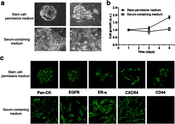
Morphological appearance, proliferation and phenotyiping of CMC cultures. a) Morphological changes of floating mammospheres/clusters in stem cell-permissive medium (upper panels) from a cobblestone-like morphology to spindle-like cells in adherent monolayers (lower panels), after differentiation for 15 days in serum-containing medium. Phase-contrast images, original magnification 10X. b) Representative growth curves of CMC cells selected under stem cell-permissive or differentiation medium, showing the in vitro proliferative potential of cultures. Arbitrary units (a.u.) are referred to the number of living cells at day 1. Data represent the mean ± s.e.m. c) Enrichment in CD44+ cells and marker expression profile of CMC cells grown in stem cell-permissive or differentiation medium. Immunofluorescence analysis of pan-cytokeratin (Pan-CK), EGFR, ER-α, CXCR4 and CD44 in CMC spheroids (upper panels) and after 15-day exposure to serum-containing medium. Images from confocal microscopy, original magnification 100X.
