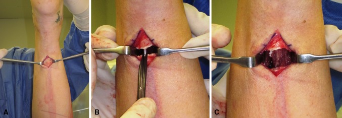
Fig. 2A–C.

The GSR surgical technique is illustrated. (A) An incision is made at the middistal 1/3 of the posterior leg. The leg is elevated and the heel is at the top of the photograph. Note the white gastrocnemius fascia. (B) After incision of the gastrocsoleus fascia, the medial raphe is seen. The raphe is about to be cut with scissor. (C) Note separation of fascia and underlying muscle.
