Fig. 1.

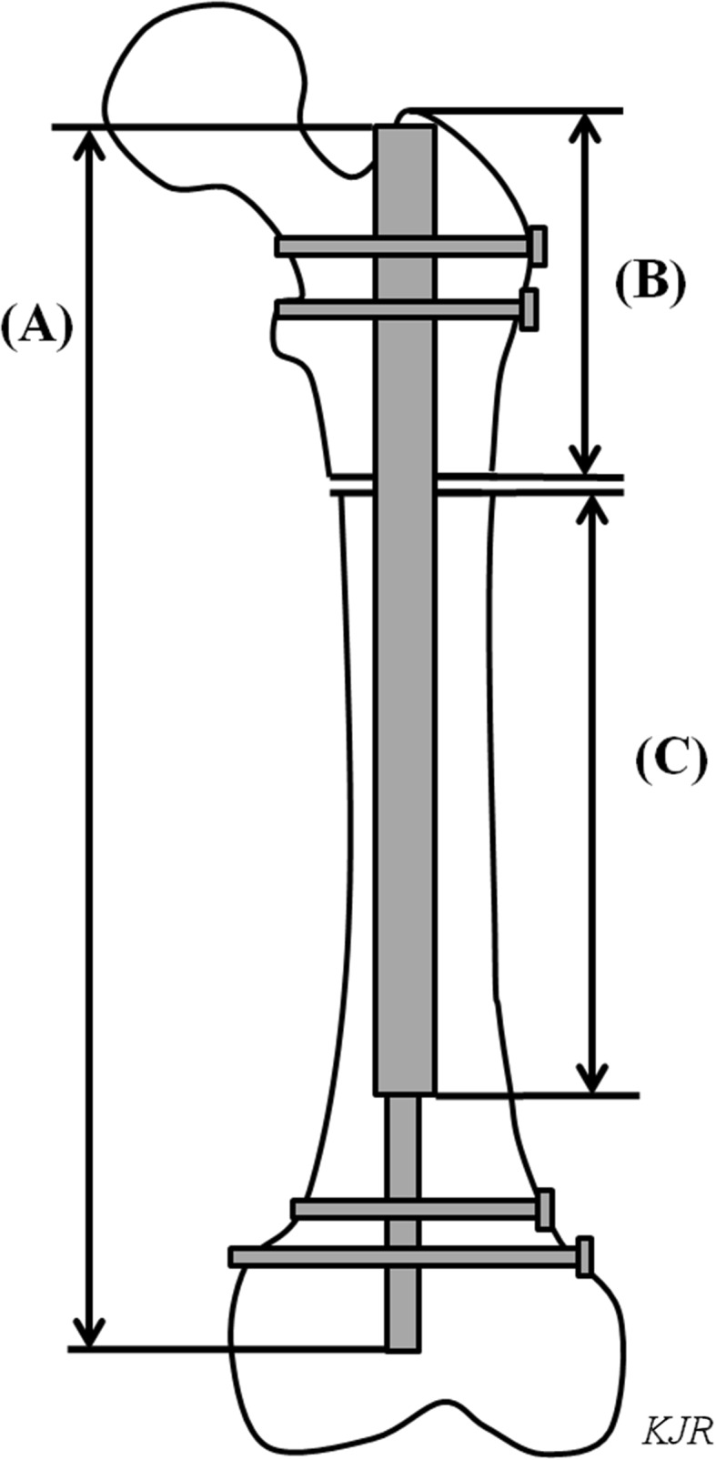
A schematic image of the left femur with the ISKD device inside shows the total length of the nail (A), the length of the proximal fragment (B), and the length of the thicker portion of the nail within the distal fragment (C).

A schematic image of the left femur with the ISKD device inside shows the total length of the nail (A), the length of the proximal fragment (B), and the length of the thicker portion of the nail within the distal fragment (C).