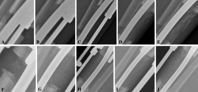
Fig. 4A–J.
Serial lateral plain radiographs show a tibia undergoing lengthening in (A–E) the treatment group and (F–J) the control group. Radiographs are from (A, F) 1 month to (E, J) 5 months postoperatively with 1-month increments between each radiograph. More callus regeneration is seen in the treatment group.

