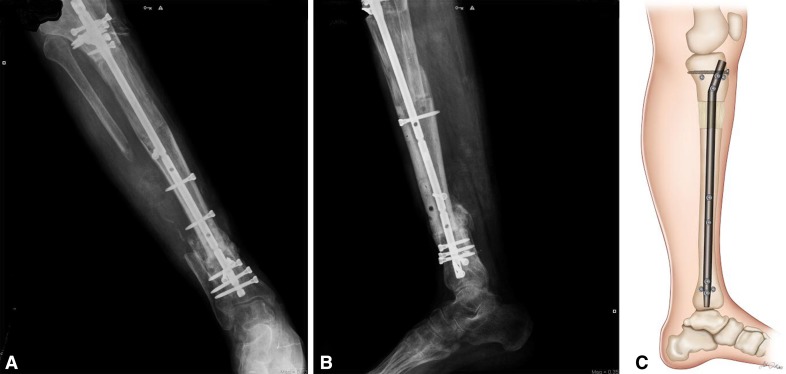
Fig. 6A–C.
Radiographic images and drawing of the tibia at 5 months followup showing removal of the circular external fixator; the proximal holes of the nail were locked with a free-hand technique. (A) AP radiograph showing the consolidated lengthening regenerate, the nail is locked proximally, and union at the docking site. (B) Lateral radiograph showing the consolidated lengthening regenerate, the nail is locked proximally, and union at the docking site. (C) Drawing of a lateral tibia image showing union at the docking site, the nail locked proximally, and the consolidated lengthening regenerate.

