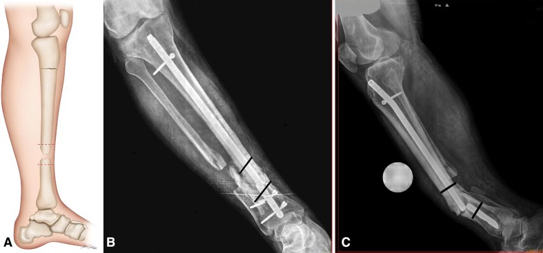
Fig. 7A–C.
Drawing and radiographs of a tibia are shown indicating the margins of resection as determined on AP and lateral radiographs. (A) Drawing of a lateral tibia showing the margins of resection (red dotted lines). (B) A 57-year-old man (Patient 4) who had tibial nonunion after three unsuccessful operations resulting from an open tibial fracture. AP radiograph of the right tibia showing a broken intramedullary nail and margin of the resection (black lines). (C) Lateral radiograph of the right tibia showing a broken intramedullary nail and margin of the resection (black lines).

