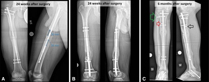
Fig. 5A–C.
AP (left) and lateral (right) radiographs show distraction measurements and consolidating bone regenerate in selected representative case examples. (A) A 14-year-old boy underwent anterograde femoral lengthening for a 3.8-cm congenital leg length discrepancy and 20° external rotation deformity. (B) A 30-year-old man underwent retrograde femoral lengthening for a 3.6-cm leg length discrepancy, 7° genu valgum, and 10° external rotation deformity due to posttraumatic growth arrest. (C) A 41-year-old man underwent tibial lengthening for 4.0-cm shortening from bone loss and tibiotalocalcaneal arthrodesis. Radiographs also show additional screw stabilization across the proximal tibia-fibula joint (green arrow), a lateral blocking screw to prevent valgus malalignment (red arrow), and a posterior blocking screw to prevent procurvatum deformity (black arrow).

