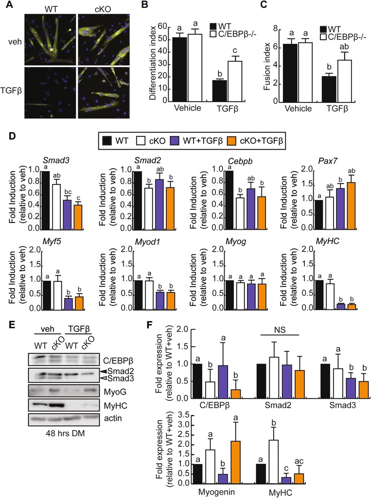
Figure 5.

Inhibition of myogenesis by TGFβ is partially rescued by loss of C/EBPβ expression. (A) Representative images of myosin heavy chain expression by immunocytochemistry in primary myoblasts isolated from cKO or control (WT) mouse hindlimb and induced to differentiate in low serum for 48 h in the absence (veh) or presence of TGFβ treatment. (B) Differentiation indices (#myonuclei/#total nuclei) of cells cultured and treated as in (A). Means marked by different letters are statistically different from one another, with a minimum of P < 0.05, n = 3. Error bars represent the SEM (C) Fusion indices (#myonuclei/#myotubes) of cells cultured and treated as in (A). Counts exclude mononucleated myosin heavy chain positive cells. Error bars represent the SEM; means marked by different letters are statistically different from one another, meeting a minimum cutoff of P < 0.05, n = 3. (D) RT-qPCR analysis of Smad2, Smad3, Cebpb, Pax7, and myogenic marker expression in primary myoblasts differentiated as in (A). Means with different letters are significantly different from one another with a P < 0.05, n ≥ 3. (E) Representative western blots of C/EBPβ, Smad2, Smad3, myogenin, and myosin heavy chain in primary myoblasts differentiated as in (A). Means with different letters are significantly different from one another with a P < 0.05, n = 5. (F) Quantification of C/EBPβ, Smad2, Smad3, myogenin, and myosin heavy chain protein expression from (E) relative to vehicle-treated WT control cells. Error bars are the SEM. Means marked with different letters are statistically different from one another, meeting a minimum cutoff of P < 0.05, n ≥ 3.
