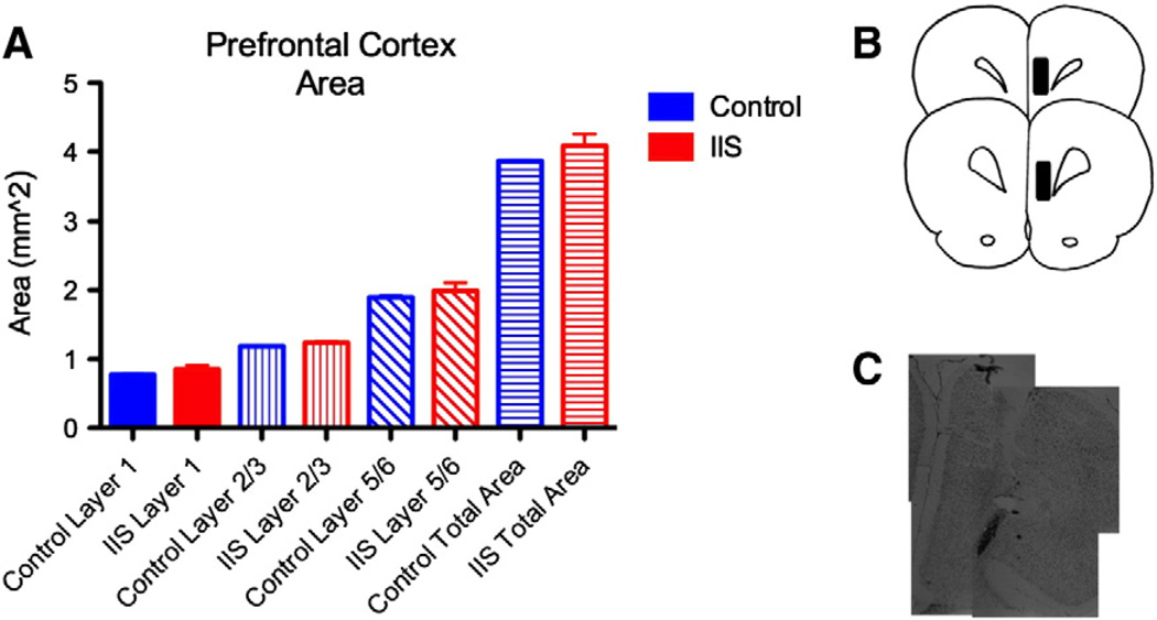
Fig. 5.
PFC structure is unaltered after early life IIS. We measured layer areas in histological sections from animals that had experienced early life IIS, littermate controls. There were no alterations in the areas of any of the layers of the PFC after early life IIS (A). (B) Shows the location of the injection site in the cohort. (C) shows thionin stained images of the electrode track lesion taken at p65.

