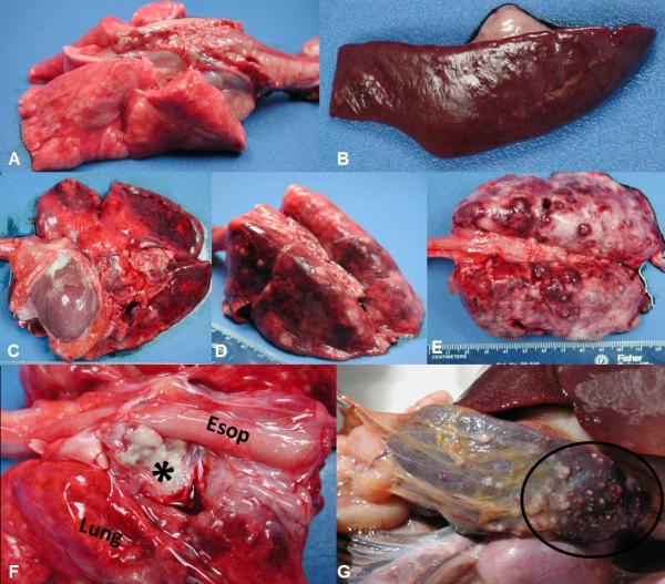
Figure 5.
Macroscopic findings in the lung, lymph node, and spleen of NHPs challenged by aerosol with F. tularensis. (A) Normal lung, non-challenged CM. (B) Normal spleen, non-challenged RM. (C) Lung, AGM 2 (D) Lung, CM 3, and (E) Lung, RM 2,. Hemorrhagic, necrotizing, and/or pyogranulomatous foci on the pleural surface with congestion and edema and fibrinous pleuritis (most noticeable in the RM); note the failure of lung lobes to collapse. (F) Tracheobronchial lymph node, CM 2. The lymph node is effaced by tannish-white caseous material (*). (G) Spleen, CM2. A myriad of multifocal to coalescing, pale white, raised or flattened necrotic foci on the capsular surface. Esop = esophagus.

