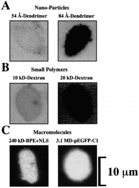
Fig. 2.

A–C Fluorescence microscopy analysis of the transport properties of isolated adult cardiomyocyte nuclei. A NPC sieving properties were studied with FITC-labeled dendrimers of 5.4 nm and 8.4 nm (54 and 84 Å) diameter. All nuclei allowed the entry of 5.4-nm diameter (54 Å) and almost all excluded the 8.4-nm-diameter (84 Å) dendrimer. B NPC sieving properties were also studied with conventional FITC-labeled dextrans of 4–150 kDa. The cutoff point for sieving was near 20 kDa as most nuclei excluded this probe. C Macromolecular import and transcriptional-translational capacity of the nuclei were tested with nuclear-targeted B-phycoerythrin (BPE+NLS) and with the plasmid for enhanced green fluorescence protein pEGFP-C1. The light detected from nuclei incubated with pEGFP-C1 comes from the expressed pEGFP product, EGFP. All probes were applied to the nuclear bath and were left throughout the imaging, as this did not interfere with the observation. Some images (e.g., 20 kDa dextran, B) are noisy due to the high gain of the photomultiplier required to detect the signal without increasing the intensity of the laser beam, which would otherwise harm the preparation
