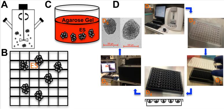
Figure 2. Multicellular tumor spheroids (MCTS) in vitro production techniques of ES.
(A) Spinner flask spheroid cultures. (B) Micro-etched Nano-cultures. (C) Biologically (e.g. Collagen gel) derived 3D matrices cultures. (D) ES Hanging-drop cultures. (D1) ES cell line counting using Beckman Coulter Vi-Cell XR Cell Viability Analyzer. (D2) 20μL of cell suspension (100 ES cells) plated on the lid of a Greiner 96-well plate. (D3) The lid was placed back on the 96-well plate containing 100 mL of RPMI (cell culture complete medium) and carefully placed in the incubator for 72 hours. (D4) The lids were removed and 300 μL of RPMI was added to a Nano-Culture® Plate (Scivax NCP-LS) to allow the drop to come in contact with the media, re-incubate for one hour and remove 100 μL. (D5) Spheroids were imaged using the GE InCell Analyzer 6000. (D6) Images of ES spheroids cells at 2 × 10⁴ cells / mL and 5 × 10⁴ cells /mL.

