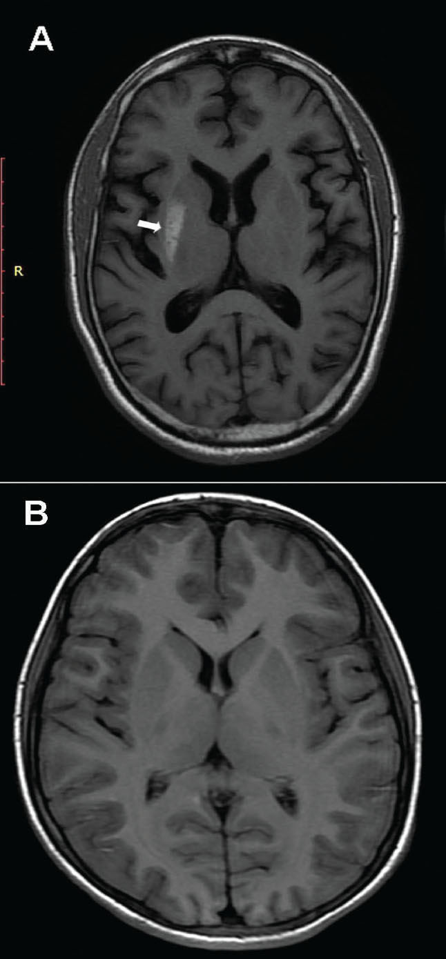
FIGURE 1.

(A) An MRI T1-weighted axial image showing an irregular area of high signal intensity involving the posterior right putamen without any appreciable adjacent edema or mass effect. (B) A 6-month re-scan shows complete disappearance of the putaminal lesion.
