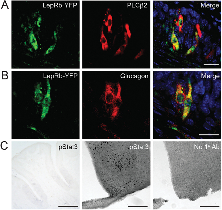
Figure 1.
LepRb is expressed, but does not activate STAT3, in taste cells. (A, B) Colocalization of LepRb-driven YFP (green, using anti-GFP antibody) and Type 2 taste cell markers (A) PLCβ2 and (B) glucagon in taste cells of mouse vallate papillae. Blue, DAPI. Scale bars, 20 μm. (C) Phosphorylated STAT3 (pSTAT3) immunostaining in mouse tissues after leptin treatment (5 μg/g; i.p. injection 45min prior to euthanasia): vallate papillae (left; scale bar, 125 μm); hypothalamus (middle; scale bar, 250 μm); hypothalamus with no primary antibody (right; scale bar, 250 μm). DAPI, 4′,6-diamidino-2-phenylindole.

